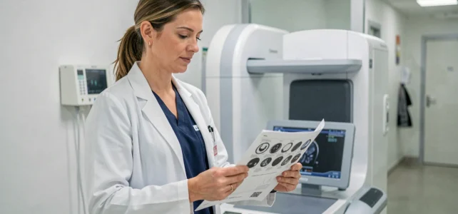
Bone densitometry measures bone mineral density using dual-energy X-ray absorptiometry, a technique referred to by the acronym DEXA (Dual-Energy X-ray Absorptiometry). In Aix-en-Provence, several medical imaging centers perform this examination on the lumbar spine and proximal femur to assess the fracture risk related to osteoporosis. Recent developments in this technique go beyond simple density measurement and open up finer diagnostic perspectives.
3D-DXA Technology and Cortical-Trabecular Analysis: What’s Changing in Bone Measurement
Classic DEXA produces a two-dimensional image and calculates an overall T-score. This approach, reliable for screening, does not distinguish between the density of cortical bone (the hard outer layer) and that of trabecular bone (the inner spongy structure). However, these two compartments do not respond the same way to bone loss or treatments.
Manufacturers like DMS Imaging, a French company, now offer densitometers that include a 3D-DXA option. This technology transforms a standard DEXA acquisition of the hip into a three-dimensional model of the proximal femur, with separate analysis of cortical and trabecular density. The examination time for the patient remains the same as that of a conventional DEXA.
This approach has been validated against quantitative computed tomography (QCT) in multicenter studies. It is still rarely mentioned on the websites of regional imaging centers, even though it represents a significant qualitative leap. An article dedicated to bone densitometry in Aix en Provence on Smart Mag details the dynamics of the local health hub around these innovations.
The clinical interest is direct: differentiating cortical loss from trabecular loss helps the rheumatologist tailor treatment. A patient with marked trabecular erosion but preserved cortical bone does not require the same therapeutic strategy as a patient whose cortical bone is thinning.
Opportunistic Screening via Chest CT and Artificial Intelligence
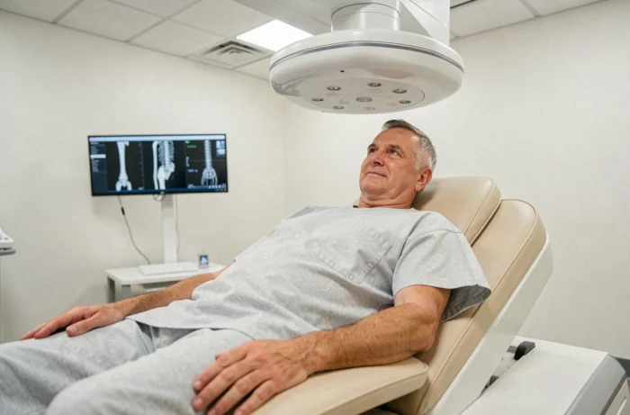
A recent research avenue involves leveraging chest CT scans performed for other reasons (pulmonary assessment, oncological follow-up) to estimate vertebral bone density. This is referred to as opportunistic osteoporosis screening.
Studies published in 2026 and reported by the RSNA (Radiological Society of North America) show that an automated chest CT can reliably measure bone density without additional acquisition or extra radiation. Artificial intelligence algorithms analyze the visible vertebrae on the scan and produce an estimation of fracture risk.
For medical imaging centers equipped with scanners, this approach could help identify at-risk patients who would never have been referred for dedicated bone densitometry. The National Osteoporosis Foundation (NOF) dedicated a session to AI applied to bone health at its 2026 conference, indicating that the topic has moved beyond the experimental stage.
Current Limitations of Opportunistic Screening
This type of screening does not replace DEXA for longitudinal monitoring. DEXA remains the reference examination for comparing two measurements over time, provided they are performed on the same device and according to an identical protocol. Opportunistic screening via CT serves as a filter: it detects but does not monitor.
Reimbursement and Prevention Pathways: The Updated French Framework
Bone densitometry is coded PAQK007 in the CCAM nomenclature. Its reimbursement by health insurance remains contingent on specific indications, which the websites of imaging centers rarely detail beyond a generic list.
Situations that qualify for reimbursement include:
- A personal history of fragility fracture (vertebra, femoral neck, wrist) without severe trauma
- Prolonged corticosteroid therapy or a treatment inducing bone loss (aromatase inhibitors, GnRH analogs)
- Pathologies associated with demineralization: untreated hyperthyroidism, hyperparathyroidism, hypogonadism
- A low body mass index or family history of femoral neck fracture in a first-degree relative
Outside of these indications, the examination remains the patient’s responsibility. Confusion is common: many postmenopausal women believe that DEXA is systematically reimbursed, which is not the case without documented risk factors.
My Prevention Assessment and Osteoporosis
The updated prevention pathways in France under the “My Prevention Assessment” program formally include osteoporosis as a theme, mentioning bone densitometry in case of risk factors, particularly around the age of sixty for women, according to the recommendations of the GRIO (Research and Information Group on Osteoporosis). This program expands detection to the primary care physician, beyond just rheumatology or gynecology pathways.
DEXA and Body Composition: An Expanding Use
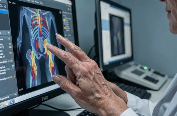
DEXA is no longer limited to bone density. Recent densitometers allow for a comprehensive body composition analysis: fat mass, lean mass, and visceral fat, with greater accuracy than traditional bioelectrical impedance.
This use is of interest in sports medicine, nutritional follow-up, and the management of sarcopenia (age-related muscle loss). In Aix-en-Provence, where the medical imaging offering is expanding, this dual function of the same device represents a lever for optimization for centers investing in state-of-the-art densitometers.
The body composition measured by DEXA also serves as a reference in clinical research protocols. When a therapeutic trial needs to quantify changes in muscle mass under treatment, DEXA serves as the gold standard, ahead of CT and MRI for this type of segmental measurement.
Bone densitometry in Aix-en-Provence is part of a dynamic where DEXA technology exceeds its initial role of osteoporosis screening. Between 3D analysis of the femur, opportunistic screening via AI on chest CT, and body composition measurement, imaging centers have tools whose diagnostic potential remains largely underutilized in everyday practice.