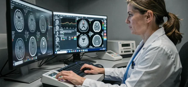
Every year in France, millions of imaging exams produce large files, encoded in technical formats, that must be transferred between the radiologist, the primary care physician, the specialist, and sometimes the patient themselves. This journey, long fragmented between CD-ROMs and postal mail, increasingly relies on specialized digital platforms. Understanding how these tools work and where their limits lie allows for a better grasp of the ongoing transformation.
The DICOM format, the technical foundation of the medical imaging journey
Before discussing platforms, it is essential to understand what circulates. Medical images (X-rays, MRIs, CT scans, ultrasounds) are not just simple JPEG files. They are produced in DICOM format (Digital Imaging and Communications in Medicine), an international standard that encapsulates both the image and clinical metadata: patient identity, exam date, acquisition parameters, modality type.
This format ensures that any compatible software can read and display the exam without loss of diagnostic information. However, it imposes constraints. DICOM files are large, sometimes several hundred megabytes for a single series of MRIs. Their transmission requires suitable network infrastructures and appropriately sized storage systems.
The PACS (Picture Archiving and Communication System) is the server that archives and distributes these images within a healthcare facility. It receives images from the scanner or MRI, indexes them, and makes them available to physicians via an integrated viewer. When a radiologist describes the website irld.net in detail, they specifically mention this connection between image production and archiving systems that conditions the entire subsequent journey.
Imaging sharing platforms: how images leave the hospital
The PACS works well within a facility. The difficulty arises when an exam needs to be consulted by an external professional, during a tele-expertise, a multidisciplinary consultation meeting (RCP), or simply when the patient changes doctors.
Specialized platforms solve this problem by creating an exchange layer above local PACS. Their operation follows a common logic:
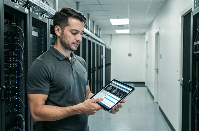
- The PACS of the producing facility pushes the images to the platform via a secure gateway, without the radiologist needing to intervene manually for each transfer.
- The platform indexes the exam, associates it with the patient file, and makes it accessible to authorized healthcare professionals, regardless of their location.
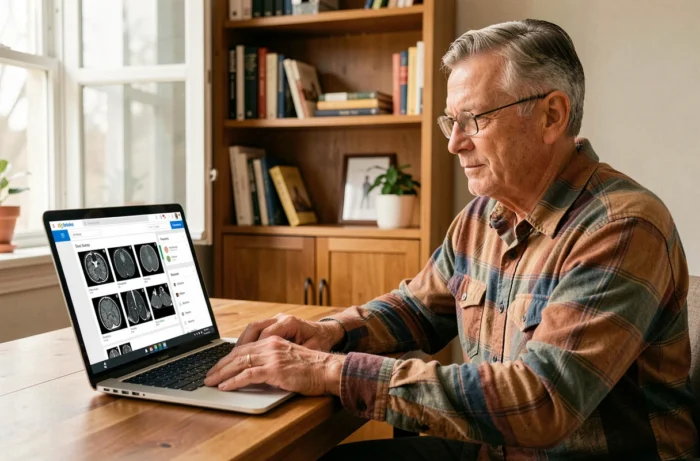
- The receiving physician accesses the images via a web viewer, without needing to install specific software or receive a CD-ROM by mail.
This model is based on a principle of data federation rather than centralization. The images remain stored on the original PACS or a regional node. The platform merely orchestrates access.
The DRIM-M project and the national scale
The DRIM-M project (Data Radiology Medical Imaging and Nuclear Medicine), supported within the framework of the Ségur du numérique en santé, illustrates this logic on a large scale. Each imaging service connects its PACS to the national network via a gateway called DRIMbox. A national index of exams is integrated into the Shared Medical Record (DMP), allowing any authorized healthcare professional to find an exam conducted anywhere in the territory.
The patient does not store their images in My health space. However, they can consult them via a link integrated into the report deposited in their personal space. This architecture avoids duplicating large files while ensuring patient access.
Interoperability and regulatory framework: where the field stands
Interoperability between systems remains the main point of friction. Two facilities equipped with different PACS do not automatically communicate, even if both comply with the DICOM format. The IHE (Integrating the Healthcare Enterprise) integration profiles and the recently updated CI-SIS interoperability framework define the exchange rules. The CI-SIS notably standardizes the structure of the “imaging file” and the sharing modalities between heterogeneous systems.
Field feedback varies on the actual level of adoption. Some hospital groups have deployed operational regional solutions (like the e-mage platform in the Grand Est region). Other territories remain at a very partial connection stage, with private radiology practices still dependent on physical media.
Tele-expertise and RCP as structuring use cases
Imaging platforms are not only used to “send” images. They enable precise clinical uses. In tele-expertise, a radiologist can submit a complex exam to a highly specialized colleague without the patient having to travel. In oncological RCP, all specialists access the same series of images during the case discussion.
These use cases justify the investment in platforms far beyond mere logistical convenience. The speed of access to images directly conditions the diagnostic timeline, and in certain pathologies, this timeline has a documented clinical impact.
Current limitations of medical imaging platforms
The deployment of these solutions faces several concrete obstacles:
- Updating RIS (Radiological Information System) software to versions referenced by Ségur involves significant costs and integration time for radiology structures, particularly private practices.
- The question of health data hosting requires the use of certified HDS hosts, which limits options and increases infrastructure costs.
- The ergonomics of web viewers lag behind the heavy reading stations used in diagnostic radiology. A web viewer does not replace a diagnostic console for exams requiring 3D reconstructions or fine measurements.
The available data does not yet allow for precise measurement of the national coverage rate of the DRIM-M network. Deployment progresses in successive waves, and the completion of the full network remains contingent on the effective equipping of each image-producing site.
The transformation of the medical imaging journey is as much a matter of infrastructure as it is of political will. The tools exist, the standards are defined, and the regulatory framework is refining. What still separates the current situation from a fluid and widespread sharing is less about technology and more about the speed of adoption in the field, practice by practice, service by service.